


![]()
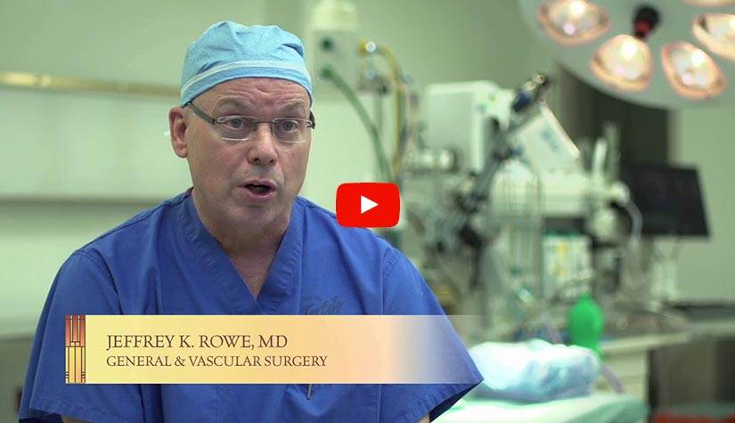
Vascular Testing
Vascular Testing at Mason City Clinic
Comprehensive vascular testing is one of many critical services we provide at Mason City Clinic. Our vascular testing currently includes:
Contact our Vascular Surgery Center: 641.494.5261
Abdominal Aortic Aneurysm (AAA) Ultrasound
 The aorta is the body’s largest blood vessel. It starts in the heart and continues through the chest and abdomen to the legs. An aneurysm is a bulge in the artery wall when the walls balloon outward with the pressure of the blood flow inside. If an aneurysm bursts, it can cause heavy, life-threatening internal bleeding. Aneurysms can form in arteries throughout the body, but most occur in the aorta. If the aneurysm occurs in the lower section of the aorta, it is known as an abdominal aortic aneurysm; if it occurs in the portion of the aorta in your chest, it is called a thoracic aortic aneurysm.
If an abdominal aortic aneurysm is suspected, an aortic ultrasound will be ordered to confirm this diagnosis. Ultrasound utilizes sound waves in scanning the aorta to give detailed information concerning the blood flow and quality of musculature around the area. The procedure requires a soft gel to be spread across the area to be observed, which helps the sound waves travel between the machine and the body. The actual imaging itself is painless, but if the area to be imaged was tender beforehand, some discomfort may ensue.
If an aneurysm is discovered, it will be monitored closely through regular ultrasound scanning. Many remain small and never rupture. Large, fast-growing, leaking or painful aneurysms may require surgery. Surgery is performed immediately on aneurysms that threaten imminent rupture or that have already ruptured, although the procedure is less successful once the vessel has burst.
Patients at risk for this condition, including smokers and those over the age of 60, should be screened regularly for an abdominal aortic aneurysm through imaging tests or genetic testing.
Venous Ultrasound
Venous ultrasound is a noninvasive imaging test that uses sound waves to create pictures of vein structures and blood flow to help accurately diagnose venous conditions.
Also known as lower extremity ultrasound, this diagnostic test can assess the veins for diseases such as dilatation, known as varicose veins, that can cause pain and tiredness of the legs. It also assesses if there is a clot in the veins that could possibly lead to pulmonary embolism. Your doctor will determine which type of procedure is most effective for your individual symptoms or condition. Once the results have been analyzed, your doctor will determine a personalized treatment plan for your condition.
Arterial Doppler/Duplex Ultrasound
An arterial Doppler/duplex ultrasound is often performed to evaluate blood flow in patients complaining of leg or arm pain, numbness, tingling and fatigue, as these symptoms may be indicative of a narrowing or blockage of the major arteries. Doppler technology uses sound waves to detect blood flow and identify any differences in blood pressure within different areas of the arms and legs.
During the Doppler/duplex ultrasound procedure, blood pressure is taken with a cuff at several areas along the arm and legs, and a transducer is moved across the area to detect blood flow through the areas before and after the blood pressure cuff is inflated. Patients may experience mild cramping as the cuff cuts off circulation in the targeted area. Patients can return to their regular activities immediately after the test.
Carotid Duplex Ultrasound
 Carotid duplex ultrasound is a diagnostic procedure that uses sound waves to detect blood flow problems in the carotid arteries. The carotid arteries are located in the neck and send blood to the brain. Blood clots and narrowed arteries are among the conditions that can be diagnosed through a carotid duplex. This test is commonly performed on patients who recently had a stroke or transient ischemic attack (TIA).
Carotid duplex is usually performed with the patient lying down on their back. A gel is applied to the skin and a handheld machine known as a transducer is run lightly across the neck along the carotid arteries. The transducer sends sound waves through the neck, forming imaging of their arrangement. Patients can return to their regular activities immediately after the test.
To learn more about our vascular services, please call 641.494.5261 today or use our online form to schedule an appointment. Our vascular testing patients come to us from Albert Lea, Algona, Belmond, Britt, Buffalo Center, Charles City, Clarion, Cresco, Emmetsburg, Forest City, Garner, Greene, Hampton, Iowa Falls, Lake Mills, Mason City, New Hampton, Northwood, Osage, Waverly and nearby areas.

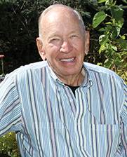 Ten years ago, my Mason City Clinic heart surgeon removed my expiration date
 Dean Haugen
Cardiology Patient
I used to visit the ER often with undiagnosed heart problems. During my last visit in 2004, they told me I probably wouldn’t make it another year. That’s when my doctor at Mason City Clinic found my mitral valve malfunction and surgically repaired it. I recently had my annual EKG exam and my cardiologist found no heart problems whatsoever. Which means I can keep raising my squash, musk melon and tomatoes.
Read More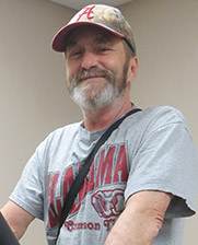 He’s the best doctor I’ve ever had – and I’ve had a lot
 Jansen Wyatt
Cardiology Patient
With no warning signs, Jansen Wyatt, 54, suddenly collapsed in his home last November. His daughter-in-law rushed him to the ER at Palo Alto County Health System in Emmetsburg, where they diagnosed a severe heart attack. Jansen was helicoptered to Mason City’s Mercy Medical Center. Samuel Congello, DO, a board-certified cardiologist with Mercy’s Heart and Vascular Institute.
Read More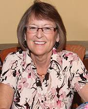 How do you handle kids if you can’t use your hands?
 Sandy Wagner
Plastic Surgery Patient
After years as a special education teacher handling children with significant disabilities and behavioral challenges, I began having limited use of my hands due to severe arthritis pain. I found myself fighting back tears every day because the pain was so bad. I could no longer enjoy one of my favorite hobbies, quilting, either. I met with Dr. René Recinos, a plastic surgeon from Mason City Clinic and recommended a procedure called arthrodesis.
Read More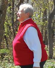 By replacing her arthritic hip and knee, Charlene can walk in the woods again without pain
 Charlene Hanson
Orthopedics Patient
Retired school teacher Charlene Hanson used to love walking in the woods – until the pain and inflammation of arthritis took it away from her. Since she had heard such great things at church and in the community about Mason City Clinic’s Orthopedic Department and Dr. Darron Jones, she made them her choice to replace her arthritic hip and knee.
Read More Getting The Hip Replacement Surgery Done At 60 Was The Best Decision I Made
 Roger Kuck
Orthopedics Patient
Roger was always very active, but a few years ago his hip started to bother him. When he would go to bed at night the pain was very severe. “I would have to lay on the floor and put my legs up on the couch to relieve the pain,” said Roger.
His orthopedic surgeon at the Mason City Clinic Dr. Darron Jones said, “I can give you cortisone shots, but this is a quality of life question.
 I trusted him 100 percent
 Donna Drake
Plastic Surgery Patient
Donna Drake lived with a growing basal cell skin cancer (the most common skin cancer) on her lower eyelid for three years. A family practitioner at Franklin General Hospital recommended Mason City Clinic’s plastic and reconstructive surgeon, Dr. Mark Mulkey, who identified the cancer and performed the extremely delicate eyelid surgery to remove it and reconstruct her eyelid.
Read More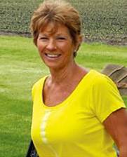 Bunion pain stopped Melody in her tracks
 Melody Wagner
Podiatry Patient
Melody Wagner loved walking until severe pain from bunions stopped her cold. She found a board-certified podiatrist at Mason City Clinic’s Podiatry Department. Podiatrists perform leading-edge surgery for bunions and hammertoes, as well as advanced treatments for diabetic feet, heel pain, ingrown toenails and more. Melody was afraid of a painful procedure and a long recovery.
Read More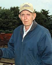 I didn’t know the pain in my leg came from a ruptured blood vessel in my chest
 Lyle Miller
Cardiology Patient
One snowy night about seven years ago, I felt a sudden pain in the back of my leg. I thought I could shake it off and went out to shovel some snow. After a few minutes, I went back inside and collapsed. My wife called 911 but the ambulance couldn’t get to me because of the snow. My neighbors used snow blowers to clear the road and my driveway so the ambulance could get to me.
Read More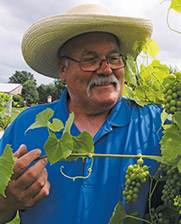 No more sour grapes about knee pain for me
 Gordon Lubbers
Orthopedics Patient
I injured my knee as a teenager and had surgery. Then, as I got older, the pain returned and began affecting my routine and overall enjoyment of life. My doctor referred me to Darron Jones, MD, an orthopedic surgeon. After a thorough exam, he said I was a candidate for knee replacement. I had the procedure in late January 2014 and was back among the grapes by the beginning of March.
Read More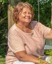 I Wanted To Dance Up A Storm At My Two Daughters Weddings, And I Did!
 Sheryl Borcherding
Orthopedics Patient
We have a lot of flower gardens on our acreage, and I was having problems kneeling to weed. I couldn’t walk long distances anymore and it was painful to get in and out of the car, walk up and down the stairs. Simple everyday tasks were hard. Eventually I was limping and in a great deal of pain.” said Sheryl Borcherding of Emmons, MN.
Read More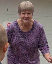 I Now Can Play Football And Basketball With My Grand Nephew.
 Jane Papouchis
Orthopedics Patient
Jane, 70, a retired nurse, is a very active person. She is a mother of one daughter and son in law, and has lots of 4 legged kids: guinea pigs, goats, Newfoundland dogs and 10 cats. I suffered with knee pain for a long time and would always take extra strength Ty-lenol.
Read More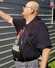 I Was Given 23 Years Of My Life…And Counting
 Gene Wagler
Cardiology Patient
Gene Wagler, a special needs teacher from Clear Lake, didn’t have a history of heart problems before his heart attack 23 years ago. Gene said, “I didn’t feel any pain that day but I had a “gnawing” sensation in my chest. Within hours Gene was in the Mercy One-North Iowa ER and Dr. Sam Congello, an interventional cardiologist.
Read More I feel great and have none of the heartburn I had for so many years and have lost 12 pounds.
 Kathleen Hanna
GERD Surgical Patient
Kathleen Hanna, 60, is a mother of four, grandmother of nine and a para-assistant at Forest City Elementary School. “I had heartburn with regurgitation for 40 years, have had an ulcer, and have been on heartburn medication for about that long too.” Having been on heartburn medication for many years her physician was becoming worried that it was affecting the function of her kidneys.
Read More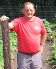 I feel great. The surgery was easy. Now I can fully enjoy my life with my wife – we garden, bike and visit with our three grown children and three grand-daughters.
 Duane Obanion
GERD Surgical Patient
In March 2019, Duane Obanion, 68, a farmer outside of Mason City, Iowa had an acid reflux attack. He aspirated into his lungs and got pneumonia. “I was in New York visiting my daughter when it happened and I ended up in the Urgent Care Center. When I got home I went to my family doctor and she put me in touch with Dr. Matthew Fabian, a general surgeon at the Mason City Clinic”.
Read More After Breaking Her Wrist In A Fall, Hampton Woman Gets Full Mobility of Her Hand And Wrist Back!
 LeAnn Strother
Plastics & Reconstructive Surgery Patient
In February 2019, LeAnn Strother 65, who is left-handed, fell and broke her left wrist. Because it was a complicated break she was referred into Dr. Rene Recinos, a plastics and reconstructive surgeon, and hand specialist at the Mason City Clinic for surgery. Most concerning for LeAnn was if this injury would impact all of the things she loves to do with her hands in the future?
Read More Dr. Scherb and his team are amazing
 Dan Rodemeyer
Orthopedic Patient
Dan Rodemeyer of Hampton, was at work when the unexpected happened. While on the loading dock, a 4,000 pound steel I-beam fell onto his foot. Dan said, “I was wearing steel toe boots, but the sheer weight of the steel beam crushed my foot and broke two of my toes. It was extremely painful to say the least.”
Dan initially went to the emergency room in Hampton. His foot had very severe soft tissue compression injuries, and there was an internal wound that was causing extreme swelling and pain. Although his big and second toe were broken, thankfully they were not compound fractures.
 Dr. Mulholland Has A Great Team And They Know Exactly What They Are Doing
 Paul Bruns
Urology (Prostate Cancer)
Paul Bruns of Clear Lake, a retired restaurateur and businessman, went to his Medicare screening with his family physician at the MercyOne Family Clinic in Clear Lake. To Paul’s surprise his physician called him back to let him know that his PSA count (protein in his prostate) was very high and that he needed to see a urologist as soon as possible.
Read More I would recommend Dr. Henrich to anyone.
 Lisa Fuller
Podiatry (Dr. Henrich)
I am back to work on my feet everyday, and I walk my dog everyday. I don’t have any pain. Lisa Fuller, a mother of three from Algona, is on her feet a lot. “I own a can redemption center with my daughter and I am walking and moving eight hours a day. I used to come home with my feet swollen red and in pain. Now when I come home, I go take my dog for a long walk,” Lisa said.
Read More Iowa Falls Woman In Full Kidney Failure, Now Planning A Hiking & Fishing Trip Out West
 Cindy Wingler
Cindy Wingler, 55, was returning from a trip out west with her boyfriend when she didn’t feel well and went to the ER at Hansen Family Hospital in Iowa Falls. She was nauseous and fatigued, and thought perhaps she was having another urinary tract infection which she had many of in her lifetime.
Read More Mason City Woman Back On Her Feet Again After Hammertoe Surgery
 Rebecca (Becky) Groh
Rebecca (Becky) Groh, 64, only goes to one podiatrist in Mason City and that is Dr. Scott Donohoe.
“I have been to Dr. Donohoe three times - for my right and left foot bunions, and hammertoes, and recently he fixed a hammertoe on my left foot that was really bothering me.
 Charles City Woman Finds Relief From Sleep Apnea Symptoms and CPAP mask with New Procedure Called Inspire Therapy
 Janet Stangl
Janet Stangl, 64, a retired administrative assistant from Charles City, was diagnosed with sleep apnea in 2006. Said Janet, "I was in the severe sleep apnea zone. I stopped breathing 30-50 times per hour during the night. I was prescribed the CPAP.
Read More Local man who struggled with the chronic symptoms of sleep apnea gets significant relief with new implantable device
 Keith Messenger
Keith Messenger, 44, a business owner in Mason City, was not getting quality sleep, and it was having an effect on his daytime productivity. “For five years or more I would crash (fall asleep) by 12 noon everyday.” Keith was diagnosed with sleep apnea; he was told that his tongue would fall back while he was sleeping blocking his airway during the night. He was prescribed the CPAP machine. Said Keith, “I tried 16 or 17 CPAP face and nose masks. The CPAP was so loud and the masks wouldn’t stay on my face. I was also prescribed a mouthpiece and that didn’t work either.”
Read More Garner woman says her ‘Tummy Tuck’ not only gives her self confidence, it’s the motivator for staying healthy.
 Renee Denny
Renee Denny, 59, of Garner, Iowa, a retired school administrator, wants to be a healthy and active grandmother one day.
“Five years ago I had gastric bypass surgery and lost 90 lbs. I started at 240 lbs. Although I was pleased with the gastric bypass results, some of my skin was loose and sagging, and was getting caught up in my belly button and creating infections,” said Renee.
 Local Woman Gets A Total Knee Replacement Enabling Her To Step Back Into The Joys Of Her Life!
 Suzanne Johnson
Suzanne Johnson, 74, of Mason City, had been struggling over the last few years to do the things she loves to do.
“I couldn’t do any type of walking. If my husband and I were going to a fair or flea market, I would always have him bring the electric scooter for me. It was limiting — not the day to day things around the house — but going out and doing the things that I love to do,” said Suzanne.
 Garner woman gets back to active life quicker after having a new & innovative bunion surgery, performed only at the Mason City Clinic.
 Jane Peterson
Jane Peterson 68, of Garner Iowa loves to golf, but it was getting harder and harder to do it with the bunions she had on each foot. “My feet would just get tired much sooner when I was golfing. And I was working full time at a factory, standing all day on a cement floor which became very painful. I just wanted to get my feet into shape by retirement so I could continue to play golf comfortably and be active with my 10 grandkids,” said Jane.
Read More Weight Loss Surgery Helped A Fort Dodge Man Lose 100 Pounds, Bring His High Blood Pressure To Normal, and Eliminate Knee Pain
 Scott Kuhlman
In 2020, Scott, 54, of Fort Dodge, at 5’8” weighed 342 lbs., his blood pressure was so high his doctor told him he was at high risk for a stroke, and he had extreme knee pain. Today Scott weighs 213 lbs., his blood pressure is normal, and all of his knee pain is gone.
Read More Mother Of Four And Special Education Teacher Loses 100 Pounds Through Gastric Bypass Surgery, And Feels Happier Than Ever
 Lisa Buss
Clear Lake resident and special education school teacher Lisa Buss, 56, reached a high of 280 pounds. Said Lisa, “When I was younger I was able to eat anything and not gain weight. Then as I got older and had my kids I started getting heavier and heavier, and it was very hard for me to lose the weight on my own.” On a 5’7” height Lisa was 150 pounds over a healthy weight.
Read More New Aortic Valve Replacement Procedure Called TAVR Giving Patients Their Quality of Life Back
 Rosalyn Barron
Rosalyn, 83, of Mason City, is a mother of three, grandmother of six, and was a regis-tered nurse at Mercy for 30 years.
“Over the last three years I was short of breath and getting bad chest pains. I had a stent placed in September 2021 for some blockage in one of my arteries, but I was still getting these worrisome symptoms,” said Rosalyn.
 Clear Lake Man Treated Successfully For Atrial Fibrillation Credits Cardiologist Dr. Michael Spooner For Managing His Care So Well
 Vern Toohey
In late 2019 Vern Toohey, 59, of Clear Lake, was having very rapid heartbeats and feeling exhausted. Said Vern, “I couldn’t walk across the house without feeling really tired.”
Read More Self-Detection Helped Mason City Woman Catch Her Breast Cancer Early Said Mason City Clinic Surgeon Caitlin Lund, DO, Was Compassionate & Kind
 Gina Chenowith
In April 2022, mother of two, grandmother of five and retired business teacher, Gina Chenoweth, 67, of Mason City, noticed a lump in her breast. “I was pre-occupied with planning a trip to Ireland and I had had a fibroid cyst for a long time so I really wasn’t concerned. But then I showed it to my sister Kim who told me I should not wait and get it looked at right away,” said Gina.
Read More Harold Lewis – Jennifer McCambridge, PA; Samuel Congello, DO – Cardiology (Stents, Defibrillator)
 Harold Lewis
Harold Lewis, 69, retired three years from the Winnebago company, a father of five and a grandfather of nine, remembers how he felt five years ago. “I was waking up at night and couldn’t breathe. I was also very tired and fatigued during the day. I went to see my family physician at MercyOne North Iowa Dr. (Phruns)? and then Dr. Brett Mulkey. They recommended that I go see Jennifer McCambridge, PA, in the cardiology department at the Mason City Clinic.” said Harold.
Read More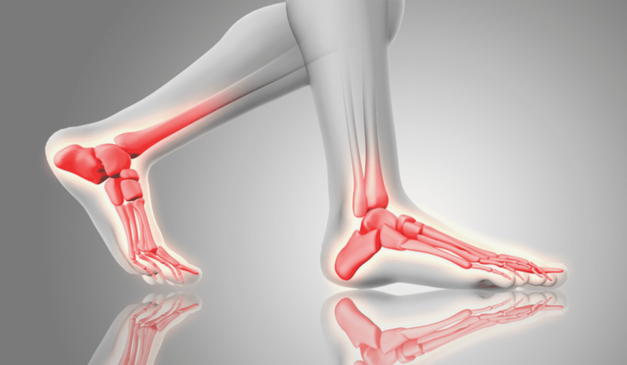 Emmetsburg Man Had Ankle Replacement & Has Increased Mobility, Less Pain Credits Orthopedic Surgeon & Podiatrist At MercyOne North Iowa Specialty Care at Mason City Clinic For His Successful Surgeries
 Larry
It was 50 years ago when Larry, 71 years old, from Emmetsburg, Iowa, was in a motorcycle accident. His ankle was crushed and his knee broken under the knee cap.
Read More Three surgeries in a year has this Woden farmer back to his daily work and golf game. Credits orthopedic surgeon Dr. Michael Crane for helping him feel ‘fantastic’
 Mike Missman
Mike Missman, 63, is a corn and soybean farmer in Woden, Iowa. It’s a physical job. “My left knee was giving me problems for many years. I felt unsteady on slippery ground in winter weather, and was in alot of
discomfort doing ‘grain climbs’ (walking up and down the stairs of the grain elevators). I was also favoring my right knee which started to bother me as well. In addition to my knees, my pinky and ring finger on my left hand and pinky and index finger on my right hand curled touching my palm. After awhile there were things that I couldn’t do with my hands
including certain mechanical functioning with tools. My grip was not tight, which also affected my golf game,” said Mike.
 George Mathews
  Clear Lake Woman Finds Joy In Outdoor Activities Again. Pain Specialist at the Mason City Clinic Helped Relieve Her Hip Arthritis Pain, Without Surgery.
 Deborah Wessels
Clear Lake resident Deborah Wessels, 69, is a proud Iowa State Cyclone alum, mother of two and grandmother of three, and enjoys being active. Deborah loves to hike, bike, golf and travel.
“For the last two years I have had a sharp pain in my left buttock. It was localized, but it started to really interfere with my activities. I used to walk three miles a day and I couldn’t do that anymore without extreme pain. I went to New York City with a college friend and we had to stop so much while walking around the city it just didn’t feel normal,” said Deborah.
 Fredericksburg Woman Gets High Quality Care Close To Home
 Elaine Westin
Mother of two and grandmother of four, Elaine Westin, 68, of Fredericksburg was struggling to do many of the activities she loves.
Said Elaine, “My husband and I went on a cruise up the east coast into Canada to see the Fall foliage and I couldn’t do many of the walking tours. My knee was weak and the pain went into my back. My flower beds suffered last summer because it was hard for me to bend over and weed. I couldn’t pick my strawberries. I even had trouble working the pedal on my sewing machine. Climbing the bleachers in the arena to watch my grandson play sports was painful and difficult. And I was not steady on my feet holding my 5 month year old grandson and I want to feel steady and sure of myself.”
 Partial Knee Replacement Allows Elma, Iowa Woman To Work and Live Without Ar-thritis Pain
 Deann Meirick
Deann Meirick, 54, of Elma, Iowa, is a housekeeper and laundry aid at a nursing home facility. She is on her feet a lot. Said Deann, “Both of my knees had been bothering me. I knew it was arthritis, but all of a sudden, my left knee was hurting to the point that I couldn’t put any weight on it.”
Read More


